My Topol fellowship problem / project:
Digital Pathology has become a rising subject within Pathology. The ability to scan slides into whole slide images (WSIs) offers many benefits to routine practice as well as research. In daily use, WSIs contribute to reduced waiting times, greater precision of diagnosis, and much less storage and handling of glass slides. Whilst these benefits have been known for some time, it is only relatively recently that the technology and the industry have developed such that slide scanners and accompanying software are now readily available for many NHS Trusts.
The implementation of Digital Pathology has gained a large traction in recent years as many Trusts seek to adopt the technology for routine use, and further integrate deep learning image analysis algorithms into standard practice. However, a natural variation has arisen in the form of intra and inter-scanner deviation. The former occurs between two scanners of the same make and model when assessing the same slide and is often negligible. The latter is often far more severe and can lead to significant change in the image quality. Differences may present in focus, colour calibration, gamma values, regions scanned, and presence of background noise. These differences can prevent full realisation of benefits for the interest of patient safety when the same slide scanned appears substantially different on two separate devices. Further, these same deviances prevent cross-Trust communication for diagnostic use and inhibits the ability to share data for research use.
This project will aim to both measure and attempt to standardise the internal and outgoing WSIs from the Royal Derby Hospital site, and if successful, attempt to apply this same method to other hospitals primarily within, and later those outside, the Trust network.
About me
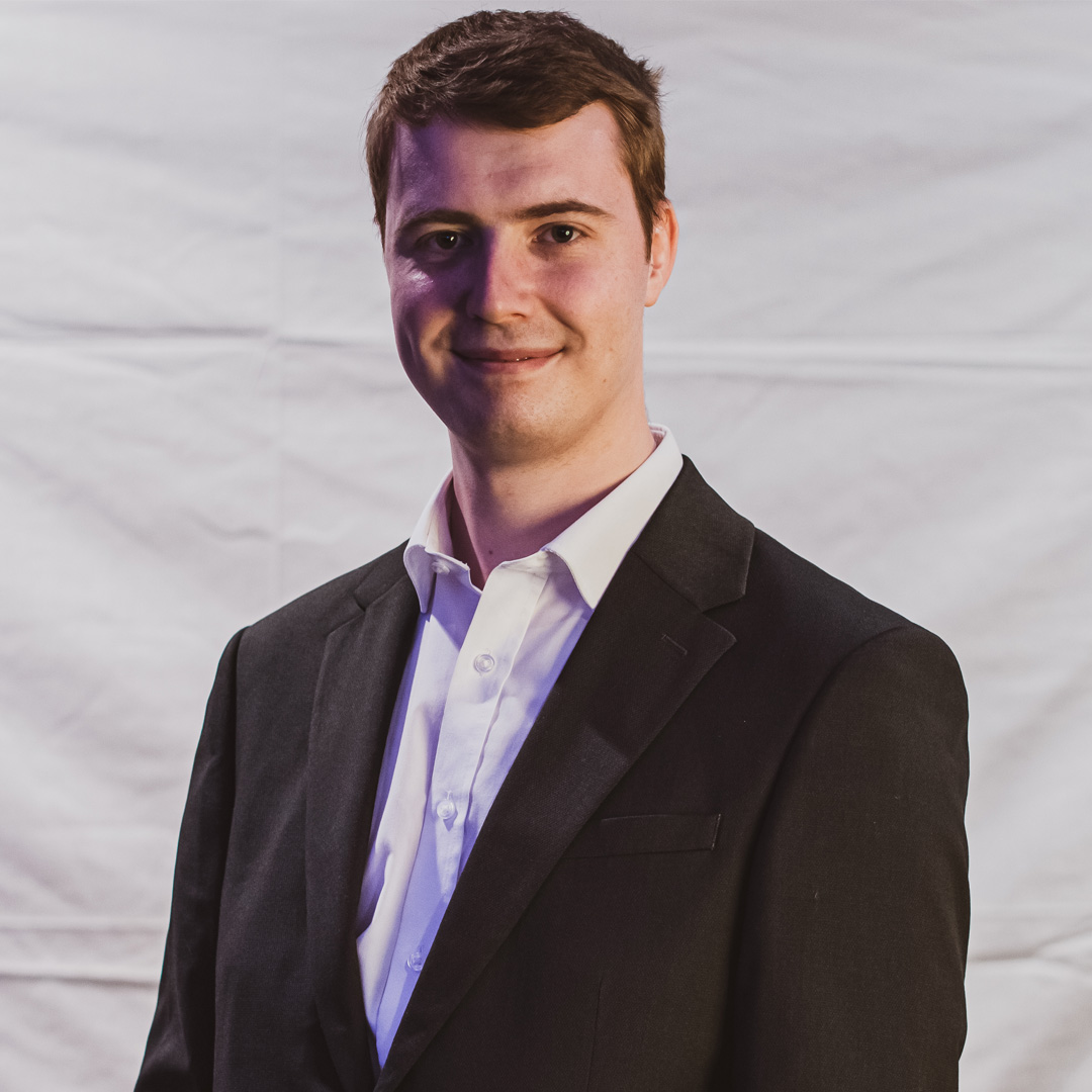
I am a Registered Biomedical Scientist within the Histopathology Department at the Royal Derby Hospital, particularly focused on Immunohistochemistry and Digital Pathology. My primary role is the implementation and validation of our laboratory’s new end-to-end Digital Pathology system, in addition to assisting in the routine operation of our Histopathology Department.
Much of my previous work has focused on the validation and integration of CE-IVD approved deep-learning image analysis algorithms to assist in quantifying ER, PR, and HER2 scoring in breast core biopsies. This work saw the production of an additional safety-net for Consultant Histopathologists, equivalent to having a 2nd pair of eyes reviewing each case. Further, this led to time savings of up to two weeks in certain patient cases, in addition to cost-savings for the care provider. The Fellowship is an opportunity to expand upon both my current and previous work, as well as to further develop my understanding of machine learning and digital health technology.

